View desktop version (interactive teaching modules and renders)

Confocal and Biological Imaging Core

Confocal, multiphoton, and enhanced-resolution light microscopy for UNLV research, with staff help on experimental design, imaging, and image analysis.

Three imaging platforms

  • Nikon A1R MP

    Multiphoton and confocal imaging with a resonant scanner, for time-lapse imaging deep within thick or living specimens.

    Details
  • Zeiss LSM 800 with Airyscan

    Confocal imaging with an Airyscan detector for enhanced resolution and signal-to-noise, suited to fixed cells, sections, and 3D stacks.

    Details
  • Leica Stellaris with white light laser

    A tunable white light laser, lifetime-based contrast with TauSense, LIGHTNING enhanced resolution, and incubation for live cells.

    Details

Start a project

Every project begins with a conversation, so the instrument and settings match your sample and question.

  1. Contact the Core

    Email andres.gutierrezgarcia@unlv.edu with a short description of your samples and what you need to see.

  2. Consultation and instrument selection

    Core staff help you plan the experiment and choose the instrument. The instrument guide is a starting point for that conversation.

  3. Training and policy agreement

    Complete training on your instrument and agree to the Core's user policies.

  4. Independent use

    Book time and image on your own. Staff-assisted imaging is also available.

Before you book

  • All microscope time must be booked in advance.
  • Training is required before independent use.
  • The Core mainly serves UNLV researchers. External users are accommodated as capacity allows.
  • Internal and external rates are available on request. See services and rates

Instruments

Three point-scanning systems with different strengths.

Nikon A1R MP

Confocal and multiphoton on one scan head, with a resonant scanner for fast time-lapse and a pulsed infrared laser for deep imaging.

Best for

  • Imaging deep in thick or scattering specimens: brain slices, intact tissue, intravital preparations
  • Fast time-lapse: the resonant scanner reaches 30 frames per second at 512 × 512 pixels
  • Long live-tissue sessions where limiting excitation to the focal plane matters (see multiphoton)
  • Second-harmonic imaging of collagen and other non-centrosymmetric structures

Not ideal for

  • Thin fixed samples where enhanced-resolution modes on the other two systems give finer detail
  • Fluorophores that need an excitation line outside the installed set
  • Experiments that need lifetime (FLIM) or spectrally tunable excitation

Configuration table: desktop version.
Zeiss LSM 800 with Airyscan

A compact confocal with sensitive GaAsP detectors and an Airyscan array detector that trades the single pinhole for 32 small ones, then reassigns their signals computationally.

Best for

  • Fixed cells and tissue sections labeled with standard 405 / 488 / 561 / 640 nm fluorophores
  • 3D stacks where resolution and signal-to-noise both matter
  • Enhanced resolution without changing the sample: Zeiss states 1.7× higher resolution in all three dimensions with Airyscan
  • Colocalization studies at the limit of light microscopy, after bead-based chromatic calibration

Not ideal for

  • Deep imaging in scattering tissue; the multiphoton system is the better tool beyond roughly 100 µm
  • Dyes that need excitation between the four laser lines
  • Long live-cell experiments

Configuration table: desktop version.
Leica Stellaris with white light laser

A spectral confocal whose white light laser can be tuned to any excitation line across the visible range, with hybrid detectors that also time each photon's arrival.

Best for

  • Multicolor experiments with overlapping spectra: tunable excitation plus adjustable spectral detection bands (see bleed-through module)
  • Live cells over hours, with stage incubation
  • Lifetime-based contrast: TauSense tools separate signals or remove autofluorescence using photon arrival times
  • Enhanced resolution with LIGHTNING adaptive deconvolution; Leica states lateral resolution "down to 120 nm"

Not ideal for

  • Deep tissue beyond the reach of single-photon excitation
  • UV-excited probes below the installed lines

Configuration table: desktop version.

Which instrument fits your sample?

Four questions. The result is a starting point for the consultation, not a booking decision.

Question 1 of 4What is the sample?
Question 2 of 4How deep do you need to image?
Question 3 of 4What matters most for the result?
Question 4 of 4Which excitation do your fluorophores need?
0 of 4 answered

Learn confocal

A summary. The interactive modules (pinhole, Airy disk, Nyquist sampling, Z-stacks, spectra, photobleaching) run in the desktop version.

1. Fluorescence

A fluorophore absorbs a photon, relaxes within picoseconds, and emits a photon of lower energy nanoseconds later: the Stokes shift lets filters separate emission from excitation. Quantum yield and lifetime describe its usefulness; the triplet state is a route to photobleaching. [Lakowicz 2006]

2. The confocal principle

Illuminate one point; detect through a pinhole in a conjugate plane; scan. Out-of-focus light arrives spread out at the pinhole and is mostly blocked, giving an optical section. PSFconfocal = PSFill × PSFdet. [Minsky 1961; Zeiss monograph 2010]

3. The pinhole

One Airy unit is the Airy disk diameter in the sample plane, 1.22 λem/NA. At 1 AU about 84 % of a point's in-focus light passes while most out-of-focus light is rejected; closing further improves resolution by up to 1.4× but discards signal, so about 1 AU is the usual compromise. [Born & Wolf; Zeiss monograph]

Reference values (point object)PH ≥ 1 AU: FWHMlat = 0.51 λexc/NA; FWHMax = 0.88 λexc/(n − √(n² − NA²)).
PH < 0.25 AU: 0.37 λ̄/NA and 0.64 λ̄/(n − √(n² − NA²)).

4. Resolution and the Airy disk

Rayleigh: two points are just resolved at 0.61 λ/NA (the Airy disk radius; the dip between them is about 74 %). Abbe: the finest transmitted grating period is λ/(2 NA). Both improve with shorter wavelength and larger NA. This site uses the Rayleigh value to define the Airy unit.

5. Pixel size and Nyquist

Sample with at least two pixels per smallest resolvable period or fine detail is aliased. Practical rule: pixel ≤ 0.5 × FWHMlat ≈ 0.25 λexc/NA (Zeiss). Strict bandwidth rule for deconvolution: λexc/(8 NA) (Scientific Volume Imaging).

6. Z-stacks

Axial resolution is roughly three times worse than lateral and scales with 1/NA². Step the focus by no more than half the optical section thickness; mind refractive-index mismatch, which stretches and blurs the stack. [Zeiss monograph; Hell et al. 1993]

7. Spectra and bleed-through

Emission spectra have red tails, so the shorter-wavelength dye leaks into the next channel. Remedies: narrower bands placed away from the overlap, sequential scanning, and linear spectral unmixing with single-label references. [Zimmermann 2005]

8. Photobleaching

Each fluorophore survives a limited number of excitations; bleaching scales with cumulative dose (intensity × time) in the linear regime. First-order decay is a planning tool; real kinetics deviate. Spend the photon budget on the frames, planes, and pixels the question needs. [Pawley 2006 ch. 39; Song et al. 1995]

9. Multiphoton excitation

Two near-infrared photons arriving together excite the fluorophore; the probability scales with intensity squared, so excitation is confined to the focus and no pinhole is needed. Infrared penetrates scattering tissue far better than visible light; bleaching is confined to the focal plane; resolution is somewhat lower. [Denk et al. 1990; Helmchen & Denk 2005]

10. Enhanced resolution

Image scanning microscopy replaces the pinhole with a detector array and reassigns each element's image (Sheppard 1988; Müller & Enderlein 2010). Airyscan is Zeiss's implementation; the manufacturer states 1.7× higher resolution in x, y, and z . LIGHTNING is Leica's adaptive deconvolution, stated to reach 120 nm laterally .

Full references are in the desktop version.

Services and rates

Instrument time, staff-assisted sessions, training, and consultation.

Instrument access

Hourly, booked in advance, after training. Independent use on all three systems.

Staff-assisted imaging

Sessions run by Core staff, for new users, difficult samples, or one-off experiments.

Training

Hands-on instrument training leading to independent use.

Consultation and analysis

Experimental design before you prepare samples; image processing and quantification after.

Access process

  • Contact the Core with a description of your samples and question (start a project).
  • Consultation and instrument selection with Core staff.
  • Training on the chosen instrument and agreement to the Core's user policies.
  • Independent use with advance booking; staff-assisted imaging on request.
  • External users are accommodated as capacity allows.

Instrument and service rates

Internal and external rates are available on request. Contact the Core for a quote for your project.

Last updated —

Hourly rates by instrument or service and user category
Instrument or serviceUnit
Loading rates…

    Core staff

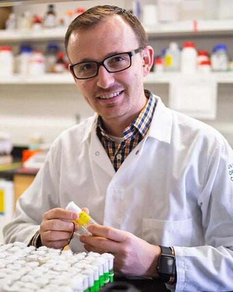
    Dr. Joseph Campanale, smiling, in a white lab coat in the laboratory, holding a sample tube

    Dr. Joseph Campanale

    Director, Confocal and Biological Imaging Core

    Dr. Campanale is an Assistant Professor in the UNLV School of Life Sciences and Director of the Confocal and Biological Imaging Core. His research uses fruit fly embryos and pancreatic organoids to understand how cells move collectively and organize into functional tissues, combining fruit fly genetics with 4‑dimensional live‑cell microscopy and high‑resolution imaging.

    He has authored numerous peer‑reviewed publications on collective cell migration, epithelial morphogenesis and live‑cell imaging methods. As Director, he oversees the facility’s scientific direction, equipment investments and upgrades, and alignment with UNLV’s research priorities.

    He helps principal investigators build robust imaging strategies into their grant proposals and mentors lab personnel in rigorous quantitative live‑cell imaging.

    Email
    joseph.campanale@unlv.edu
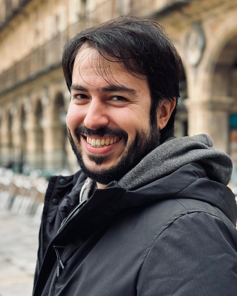
    Dr. Andrés Gutiérrez, smiling, outdoors in a winter coat

    Dr. Andrés Gutiérrez

    Manager, Confocal and Biological Imaging Core

    Dr. Gutiérrez is the Manager of the Confocal and Biological Imaging Core. He is a Biochemist (Universidad de Navarra, 2013) with a PhD in Biology (Universidad Autónoma de Madrid, 2024), and has worked with confocal microscopes and live imaging since 2014, including at the Center for Advanced Microscopy at Northwestern University’s Feinberg School of Medicine.

    His expertise covers experimental design for live and fixed samples and image analysis with tools such as FIJI/ImageJ. He has contributed to peer‑reviewed publications and methods papers on confocal and multiphoton imaging and 4D live‑cell microscopy.

    At UNLV he maintains the Nikon A1R MP, Zeiss LSM 800 with Airyscan and Leica Stellaris systems, and is the primary contact for UNLV laboratories, coordinating training, scheduling, quality control and troubleshooting.

    Email
    andres.gutierrezgarcia@unlv.edu
    Phone
    702‑774‑1550
    Office
    SEB 3253
    Facility
    SEB 1110

    Acknowledging the Core

    Please include this sentence in publications that use Core data.

    Imaging was performed at the UNLV Confocal and Biological Imaging Core.