University of Nevada, Las Vegas · College of Sciences

Confocal and Biological Imaging Core

Confocal, multiphoton, and enhanced-resolution light microscopy for UNLV research, with staff help on experimental design, imaging, and image analysis.

Instruments
3 platformsMultiphoton, Airyscan, and white-light-laser confocal
Support
Staff-assistedDesign, imaging, and analysis consultation
Access
Open to UNLVExternal users as capacity allows
Simulated Z-stack, drawn procedurally: each optical section is scanned line by line at a new depth, then the stack is tilted to show its three dimensions. Not a micrograph.
Instruments

Three platforms, one conversation

Together they cover deep-tissue multiphoton imaging, high-sensitivity and enhanced-resolution confocal imaging, and lifetime-based multicolor work on fixed and live samples.

  • Multiphoton · resonant

    Nikon A1R MP

    Multiphoton and confocal imaging with a resonant scanner, for time-lapse imaging deep within thick or living specimens such as brain slices, tissues, and intravital preparations.

    Nikon A1R MP details
  • Confocal · Airyscan

    Zeiss LSM 800 with Airyscan

    Confocal imaging with an Airyscan detector for enhanced resolution and signal-to-noise, suited to fixed cells, tissue sections, and 3D stacks.

    Zeiss LSM 800 details
  • Spectral · lifetime

    Leica Stellaris with white light laser

    A tunable white light laser, lifetime-based contrast with TauSense, LIGHTNING enhanced resolution, and incubation for live cells.

    Leica Stellaris details
Access

Start a project

Every project begins with a conversation, so the instrument and settings match your sample and question.

  1. Contact the Core

    Email andres.gutierrezgarcia@unlv.edu with a short description of your samples and what you need to see.

  2. Consultation and instrument selection

    Core staff help you plan the experiment and choose the instrument. The instrument guide is a starting point for that conversation.

  3. Training and policy agreement

    Complete training on your instrument and agree to the Core's user policies.

  4. Independent use

    Book time and image on your own. Staff-assisted imaging is also available.

Before you book

  • All microscope time must be booked in advance.
  • Training is required before independent use.
  • The Core mainly serves UNLV researchers. External users are accommodated as capacity allows.
  • Internal and external rates are available on request. See services and rates
Support

How the Core can help

Experimental design

Consultation on planning your experiment before you prepare samples: which labels, which instrument, which settings.

Staff-assisted imaging

Imaging sessions run with Core staff.

Training

Hands-on instrument training that leads to independent use.

Image analysis

Consultation on processing and quantifying your images.

People

Core staff

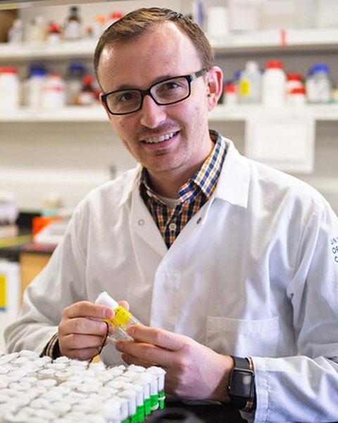
Dr. Joseph Campanale, smiling, in a white lab coat in the laboratory, holding a sample tube

Dr. Joseph Campanale

Director, Confocal and Biological Imaging Core

Dr. Campanale is an Assistant Professor in the UNLV School of Life Sciences and Director of the Confocal and Biological Imaging Core. His research uses fruit fly embryos and pancreatic organoids to understand how cells move collectively and organize into functional tissues, combining fruit fly genetics with 4‑dimensional live‑cell microscopy and high‑resolution imaging.

He has authored numerous peer‑reviewed publications on collective cell migration, epithelial morphogenesis and live‑cell imaging methods. As Director, he oversees the facility’s scientific direction, equipment investments and upgrades, and alignment with UNLV’s research priorities.

He helps principal investigators build robust imaging strategies into their grant proposals and mentors lab personnel in rigorous quantitative live‑cell imaging.
Email
joseph.campanale@unlv.edu
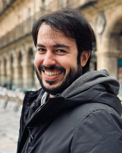
Dr. Andrés Gutiérrez, smiling, outdoors in a winter coat

Dr. Andrés Gutiérrez

Manager, Confocal and Biological Imaging Core

Dr. Gutiérrez is the Manager of the Confocal and Biological Imaging Core. He is a Biochemist (Universidad de Navarra, 2013) with a PhD in Biology (Universidad Autónoma de Madrid, 2024), and has worked with confocal microscopes and live imaging since 2014, including at the Center for Advanced Microscopy at Northwestern University’s Feinberg School of Medicine.

His expertise covers experimental design for live and fixed samples and image analysis with tools such as FIJI/ImageJ. He has contributed to peer‑reviewed publications and methods papers on confocal and multiphoton imaging and 4D live‑cell microscopy.

At UNLV he maintains the Nikon A1R MP, Zeiss LSM 800 with Airyscan and Leica Stellaris systems, and is the primary contact for UNLV laboratories, coordinating training, scheduling, quality control and troubleshooting.

Email
andres.gutierrezgarcia@unlv.edu
Phone
702‑774‑1550
Office
SEB 3253
Facility
SEB 1110

Contact and location

For urgent issues involving the core during business hours, please contact Dr. Gutiérrez or Dr. Campanale.

For emergencies, follow UNLV protocols. UNLV Police, emergency: 702‑895‑3669 from a cellphone, or 911 from a campus landline. Non‑emergency: 702‑895‑3668 (cellphone) or 311 (campus landline).

Publishing

Acknowledging the Core

Imaging at the Core has supported research projects and grant applications across the College of Sciences and allied programs. Please include this sentence in publications that use Core data.

Imaging was performed at the UNLV Confocal and Biological Imaging Core.

The multiphoton system was acquired with support from the National Science Foundation, award DBI-1726925.